
MRI or Layer x-ray
X-rays are a common method used when cancer is suspected. X-rays, also called computed tomography (DT or CT)) provide detailed images of the body's organs. The examination provides the opportunity to detect diseases in, among other things, the head, chest or stomach.
With us, you can easily book an appointment without any referral. Common questions that many patients tend to ask are, for example, questions about radiation, how the examination is carried out and how good the examination itself is?
What is the difference between MRI and X-ray?
Magnetic resonance imaging (MRI) uses magnetic fields to produce images of the body's interior. Which means that MR does not emit radiation to the patient. Computed tomography uses ionizing X-ray radiation, which can be dangerous if radiation doses are too high. The amount of radiation we are talking about is the amount we get during a year from natural radiation doses.
When examining with MR, you examine both the skeleton and the soft parts of the body, MR is an examination with a greater focus on detailed images. Magnetic resonance imaging is used, among other things, for suspected soft tissue injuries such as ligament and ligament injuries.
What can be detected with X-rays?
With X-rays, brain tumors, lung cancer, tumors in the large intestine can be detected, among other things, and it can also be seen if there is any spread to lymph nodes in the vicinity of the tumor. By finding out the cause in good time, there is a greater chance that you will receive the right treatment.
This is how the X-ray examination works
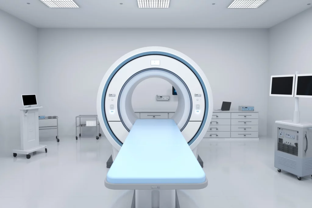
In a X-ray examination, radiation is used to produce images of the body's various organs. In order to be able to produce clearer images, you get a so-called contrast agent. The contrast agent means that the organs are seen more clearly, which means that you get clearer images. You will lie on a stretcher that is inserted into a circular X-ray machine.
The device is open at both ends, which makes it optimal for most people to perform the examination. It is important that you lie completely still while the x-ray machine takes cross-sectional images of the body. The survey takes between 10 and 15 minutes. Some may find the examination unpleasant from having to lie still for a long time, in these and other cases there are sedatives available.
What is better computed tomography or magnetic resonance imaging?
In urgent examinations, it is common to use X-rays instead of magnetic resonance imaging because the imaging takes less time. Another explanation is that X-rays are more readily available. We see big differences in the time a X-ray examination takes compared to an MRI examination.
Also big differences in how soft parts and skeleton are depicted. The difference between MR (magnetic resonance imaging) is that MR does not use X-rays like computed tomography, which means that you can examine yourself several times. Magnetic X-ray does not use any X-ray radiation, which means that you can be examined several times without any risk.
Examination of children
An X-ray examination of children can be of great benefit in being able to diagnose and treat various diseases and injuries. X-rays pass through the body during each examination, but the radiation dose is adjusted so that it is as small as possible. When examining children, you should think about preparing the child as it may feel unpleasant to have to lie in the X-ray alone for a long time. Some images may need to be retaken several times if the patient does not lie still during the examination. If necessary, sedative drugs can be given.
Pregnant women
When examining the abdomen, women of childbearing age are asked about pregnancy, as there is insufficient research into how X-rays can affect pregnant women and the fetus, women should not perform the examination. The doctor can then possibly offer other methods that are more suitable or alternatively carry out the examination after the birth.
Magnetic metal
Unlike an X-ray, no X-ray radiation is used when you are examined with a magnetic camera. Therefore, you can be examined several times without risking being affected by radiation. During an examination, it is important to empty the room of certain objects such as jewellery, bank cards etc.
This is because the objects can affect the examination in different ways. If you have magnetic metal implanted, it is not possible to perform an MRI because the magnetic field affects the magnetic rays. In those cases, other methods are usually recommended. How long the examination takes depends on the various aspects, if you use contrast media it usually takes longer. You are usually allowed to leave the hospital immediately after the examination.
Results of examination by responsible doctor within 7 days
After a magnetic camera examination without a referral at Magnetlbbaet, the scan is first reviewed by an X-ray specialist, then Magnetlabbet's doctor analyzes and compiles the results in an individual written report that you receive within 7 working days after a normal examination.
X-rays with the Magnetlab do not take long, usually 10-15 minutes. In some cases, the examination may take a little longer. You also get a USB stick from your MRI examination.